
Keratoconus |
| 09-03-2022 |
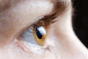 What is keratoconus?
Keratoconus is a disease of the cornea in which its central or paracentral part undergoes spontaneous, progressive thinning and bulging, eventually taking the shape of a cone.
Such reshaping of the cornea causes light rays to blur, resulting in blurred and distorted vision, which in turn hinders everyday activities such as reading or driving. It contributes to the development of irregular astigmatism.
Keratoconus usually develops in both eyes, although the progression of the changes in each eye does not have to be symmetrical. The disease usually begins in adolescence and finishes developing with the end of the body’s growth. This does not mean, however, that abnormalities in the corneal structure cannot occur at later stages in life.
Patients most frequently report to an ophthalmologist with the following symptoms:
The exact causes of keratoconus are currently unknown. One of the factors is likely to be genetics and diseases of genetic origin (such as Down syndrome).
A significant role may also be played by external factors causing eye irritation, such as mechanical injuries, caused by wrongly prescribed contact lenses or very frequent and intense eye rubbing – especially in patients suffering from atopic diseases leading to eye itchiness (e.g. allergies or eczema).
The diagnosis can be made with a slit-lamp examination and by observation of corneal thinning. If keratoconus is suspected, it is essential to perform pachymetry and corneal topography (corneal map). These examinations allow to assess the thickness of the cornea, as well as its exact shape and curvature.
The same measurements are also taken during a preliminary examination kwalifikacyjnego qualifying for a laser vision correction. They enable the ophthalmic surgeon to choose the most suitable correction method for each patient and to exclude any potential contraindications – one of which may be keratoconus.
Untreated keratoconus constitutes a contraindication to a laser vision correction procedure. Due to its progressive nature, the disease is unpredictable in terms of consequences, hence performing a laser vision correction surgery in such a case would not be safe and effective.
It has to be born in mind that using the above types of keratoconus treatment can only slow down the progression of the disease or completely stop it at a particular stage.
If keratoconus is at a more advanced stage, and continues to develop, the only effective form of treatment is a surgical procedure.
In patients suffering from keratoconus, each type of treatment increases the chance of being qualified for a laser vision correction procedure. In order to make an appointment for a preliminary qualifying examination, fill in the preliminary qualification form online or call us.
ArtLife Centrum Okulistyczne
80-398 Gdańsk
ul. Obrońców Wybrzeża 23
Centrum Horyzont
Telefon:
+48 58 557 57 77
+48 605 03 66 30
+48 506 807 111
E-mail:
Poniedziałek - piątek
9.00 - 17.00
![]()
Ta witryna używa pliki cookie. Korzystając ze strony wyrażasz zgodę na używanie plików cookie
zgodnie z aktualnymi ustawieniami przeglądarki. Możesz je w każdej chwili zmienić.
Aby dowiedzieć się więcej o plikach cookies lub je usunąć zapoznaj się z
zasadami wykorzystania plików cookies.
© 2015 - 2024 Centrum Okulistyczne ArtLife. Laserowa korekcja wzroku. Gdańsk, Sopot, Gdynia, Trójmiasto, Polska.
Design:
MK-Advice & ArtLife