Keratoconus |
| 04-07-2014 |
Keratokonus (from Greek: kerato- horn, cornea; and konos cone) – is a degenerative disorder of the eye in which structural changes within the cornea cause it to thin and change to a more conical shape than the more normal gradual curve.
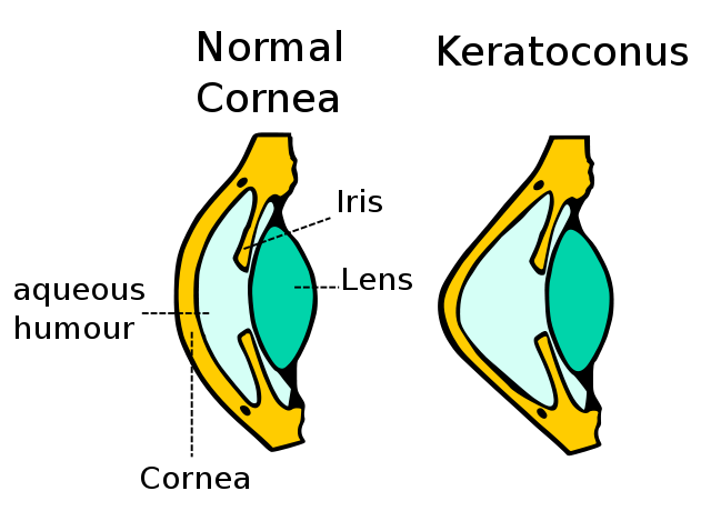
A schematic diagram showing change in cornea.
Keratoconus can cause substantial distortion of vision, with multiple images, streaking and sensitivity to light all often reported by the patient. It is typically diagnosed in the patient's adolescent years. If afflicting both eyes, the deterioration in vision can affect the patient's ability to drive a car or read normal.
SOURCE : WIKIPEDIA
Usually in early adolescence (before 18 or just after 20 years of life). In very few cases very young or mature people can also be diagnosed with keratoconus. In most cases symptoms are: slight astigmatism, blurred vision, multiple and split images. Course of the keratoconus may vary, some patients may suffer from stable form of it for many years while in other cases it may intensify very immediately (or periodically). Usually keratoconus develops for 10 to 20 years after which it stabilises.
Characteristic symptom of keratoconus is monocular polyopia which means seeing multiple ‘ghost’ images. Instead of seeing just one point, a patient with keratoconus sees many images of the point, spread out in a chaotic pattern.
Most common method allowing to regain correct vision are rigid contact lenses, known as rigid, gas-permeable, (RGP) lenses. RGP lenses provide a good level of visual correction, but do not arrest progression of the condition. Those lenses are individually designed for each patient on the basis of data from corneal topograph examination. It’s a computer-based device which precisely images the shape of the cornea in a form of a map and allows to examine various abnormalities. Lenses have to be shaped in a way that prevents pressing on the top of the cone or damaging cornea. Space between the cornea and the lens is filled with lachrymal fluid; that contributes to the improvement of the vision. Accurately chosen rigid lenses are working properly for about 18 months, after that period, due to the progression of the disease, they have to be changed. Unfortunately, in some cases, it may turn out that contact lenses themselves are insufficient. What is more, using them for a long period increases the risk of complications. That is when surgical treatment may be required. In our Ophthalmology Centre we offer contactless examinations in terms of keratoconus using a WaveLight topoliser. The examination is performed by ophthalmic surgeon.
E-mail us, make an appointment today, get yourself examined.
ArtLife Centrum Okulistyczne
80-398 Gdańsk
ul. Obrońców Wybrzeża 23
Centrum Horyzont
Telefon:
+48 58 557 57 77
+48 605 03 66 30
+48 506 807 111
E-mail:
Poniedziałek - piątek
9.00 - 17.00
![]()
Ta witryna używa pliki cookie. Korzystając ze strony wyrażasz zgodę na używanie plików cookie
zgodnie z aktualnymi ustawieniami przeglądarki. Możesz je w każdej chwili zmienić.
Aby dowiedzieć się więcej o plikach cookies lub je usunąć zapoznaj się z
zasadami wykorzystania plików cookies.
© 2015 - 2024 Centrum Okulistyczne ArtLife. Laserowa korekcja wzroku. Gdańsk, Sopot, Gdynia, Trójmiasto, Polska.
Design:
MK-Advice & ArtLife